
Research
X-Ray microtomography of bone repair within bone graft substitutes
| Principal investigator: | Karin HING |
| Co-investigator(s): | G.R. Davis and A. Parish |
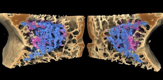
Investigation of the use of the high resolution MuCat XMT scanner which enables non-destructive visualisation of the individual struts of cancellous bone and ceramic sponge while the attenuation sensitivity enables the ceramic to be easily distinguished from the bone tissue. This technology enables medical researchers to determine in 3 dimensions the degree to which bone has grown into the ceramic, how it has been incorporated into the natural structure of the surrounding tissue and whether it has even been remodelled and replaced by new bone tissue. Further work is on-going to develop operator independant quantitative analysis.