
Research
Biosensor arrays interrogated using SPIM
| Principal investigator: | Steffi KRAUSE |
| Co-investigator(s): | Y. Zhou and J.-N. Chazalviel |
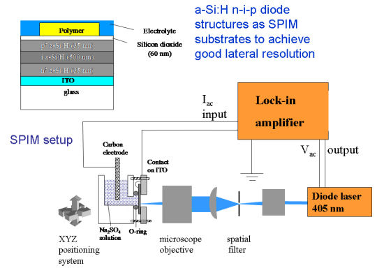 Disposable sensors based on the degradation of thin films as a result of an enzymatic reaction have been developed into efficient enzyme detectors. Film degradation has traditionally been monitored using Surface Plasmon Resonance (SPR), Quartz Crystal Microbalance (QCM) or classical ac impedance measurements. The enzyme detection principle has now been integrated with an array technology derived from a recently developed impedance imaging technique, Scanning Photo-induced Impedance Microscopy (SPIM). SPIM is based on photocurrent measurements at field-effect structures. The material under investigation is commonly deposited onto a semiconductor-insulator substrate.
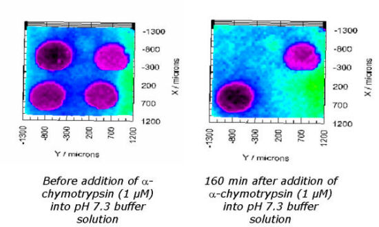 In this work, field-effect capacitors were replaced by hydrogenated amorphous silicon (a-Si:H) n-i-p photodiode structures, which have recently been shown to be suitable for SPIM measurements with good lateral resolution. To demonstrate the feasibility of SPIM for the characterization of biosensor arrays, polymer dots of the inert polymer cellulose acetate and an α-chymotrypsin sensitive poly(ester amide) were deposited onto a-Si:H n-i-p/SiO2 structures and their enzymatic degradation was monitored using a laser scanning setup.