Research
Micromechanics of mineralised biological tissues
| Principal investigator: | Andy BUSHBY |
| Co-investigator(s): | A. Boyde |
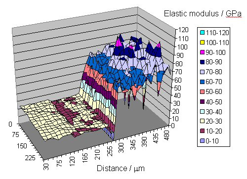
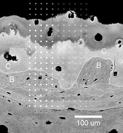 Mineralised tissues such as bones, calcified cartilage and the dental tissues (dentine, enamel and cementum) are essentially nano-composites of collagen or other proteins, mineral and water. In this project we combine nanomechanical information with quantitative microscopy to infer the composite ultrastructure of mineralised tissues and to recognise changes in those structures with disease (J. Anatomy, 203, 191 (2003)). I have recently co-organise a highly successful MRS symposium of mechanical behaviour of biological materials at the Fall Meeting, 2005. A focus issue of the Journal of Materials Research will be published in 2006 associated with the meeting.