
Research
Interaction of muscular forces with mineral nanostructure in intramembranously ossifying bones
| Principal investigator: | Himadri GUPTA |
| Co-investigator(s): | A. Karunaratne, R.V. Thakker, G.R. Davis, N.J. Terrill; and C. Esapa |
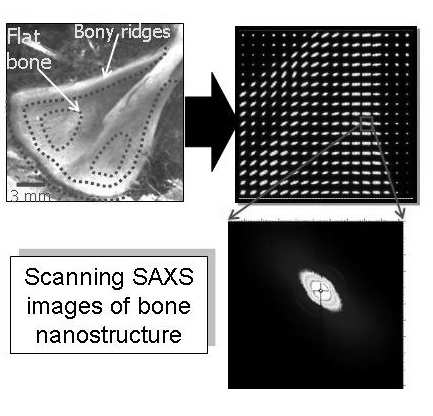 Metabolic bone disorders like rickets are associated with altered in-vivo muscular force distributions on the skeletal system. During development, these altered forces can potentially alter the spatial and temporal dynamics of mineralized tissue formation, but the exact mechanisms are not known. We use position – resolved scanning small angle X-ray scattering (sSAXS) and microCT to correlate between the nanocrystallite alignment (inside fibrils) and in-vivo muscular forces, muscle fibre diameter and muscle weight. Our model systems are the flat scapular bones of developing mice, in the intramembranous fossal regions as well as the scapular spine.